Coxa vara
Causes
Coxa vara can be present at birth (congenital) or develop over time. The causes can include:
- Developmental problems: Abnormal growth of the cartilage and bone in the femoral neck, which is the most common cause in children.
- Injury: A poorly healed fracture in the upper part of the femur can lead to the deformity.
- Metabolic bone diseases: Conditions like Paget's disease or osteogenesis imperfecta (brittle bone disease) can weaken the bone tissue.
- Infection: An infection in the bone (osteomyelitis) can cause damage and alter growth.
- Genetic disorders: Certain genetic conditions, such as multiple epiphyseal dysplasia, can lead to coxa vara.
Symptoms
Symptoms often appear as a child begins to walk, between the ages of two and six, and may include:
- A limp or waddling gait.
- One leg appearing shorter than the other.
- Pain in the hip or leg, though it can also be painless in some cases.
- Limited hip movement, especially when moving the thigh out to the side.
- In some bilateral cases, an exaggerated curve in the lower back (lumbar lordosis).
Diagnosis
A doctor typically diagnoses coxa vara using a medical history, physical examination, and imaging.
- Physical Exam: The doctor will observe the child's walking and check for limited range of motion in the hip.
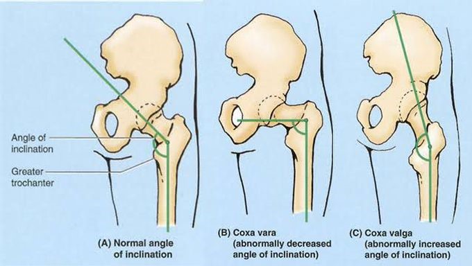
- X-rays: These are the primary tool for diagnosis and show the decreased angle between the femoral head and shaft. Advanced cases may reveal a characteristic "inverted Y-shaped" ossification defect.
- CT scan: In some cases, a CT scan may be used to get a more detailed view of the bone structure and plan for surgery.
Treatment
Treatment options depend on the severity of the condition and the patient's age.
- Observation: In mild cases, a doctor may recommend monitoring the condition with regular follow-up checks as the child grows.
- Surgery (Osteotomy): For more severe cases, a surgical procedure called a valgus derotation osteotomy is often performed. The surgeon reshapes the femur to correct the angle, reduce shearing forces, and improve hip alignment.
- Post-operative care: After surgery, the patient may need to wear a hip spica cast for a period of time to immobilize the joint while it heals.
- Physical therapy: Following surgical treatment, physical therapy can help to strengthen muscles and regain flexibility.
Do you want me to search for treatment options for a specific age group?
